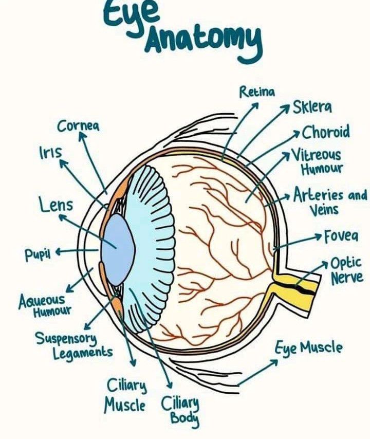
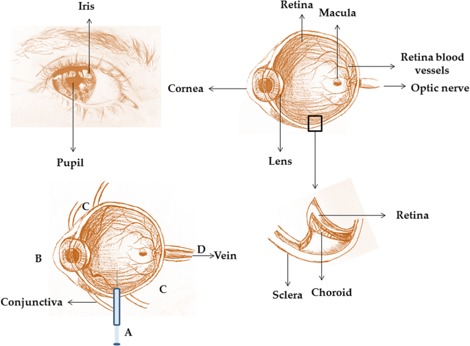
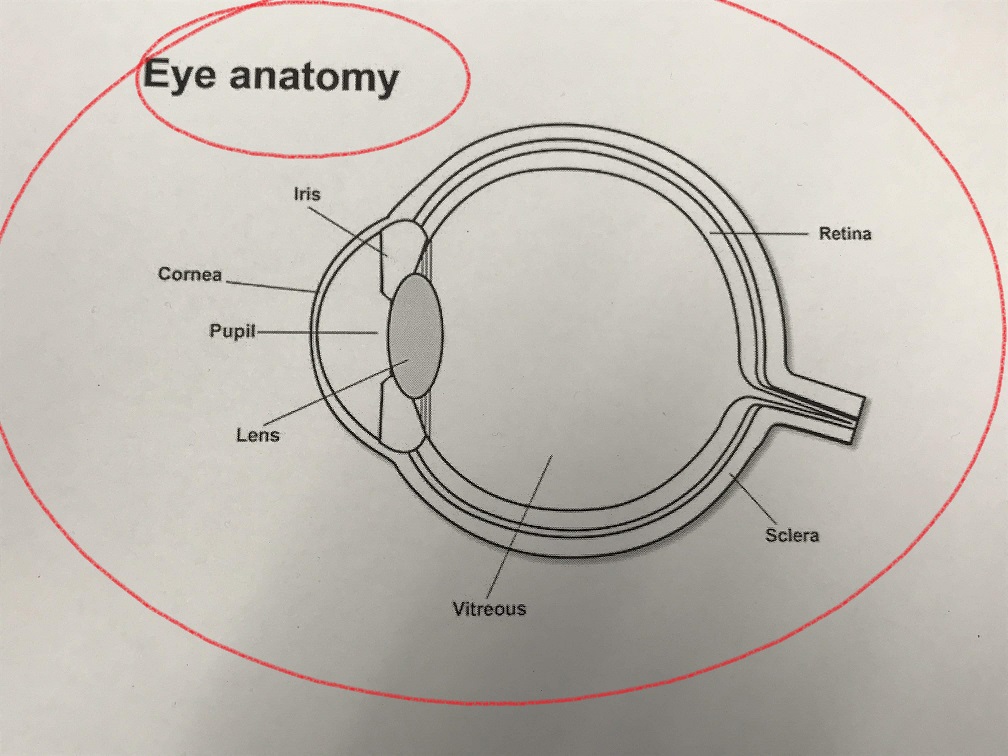
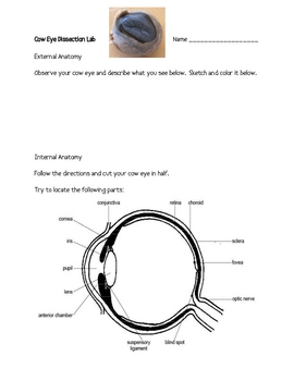
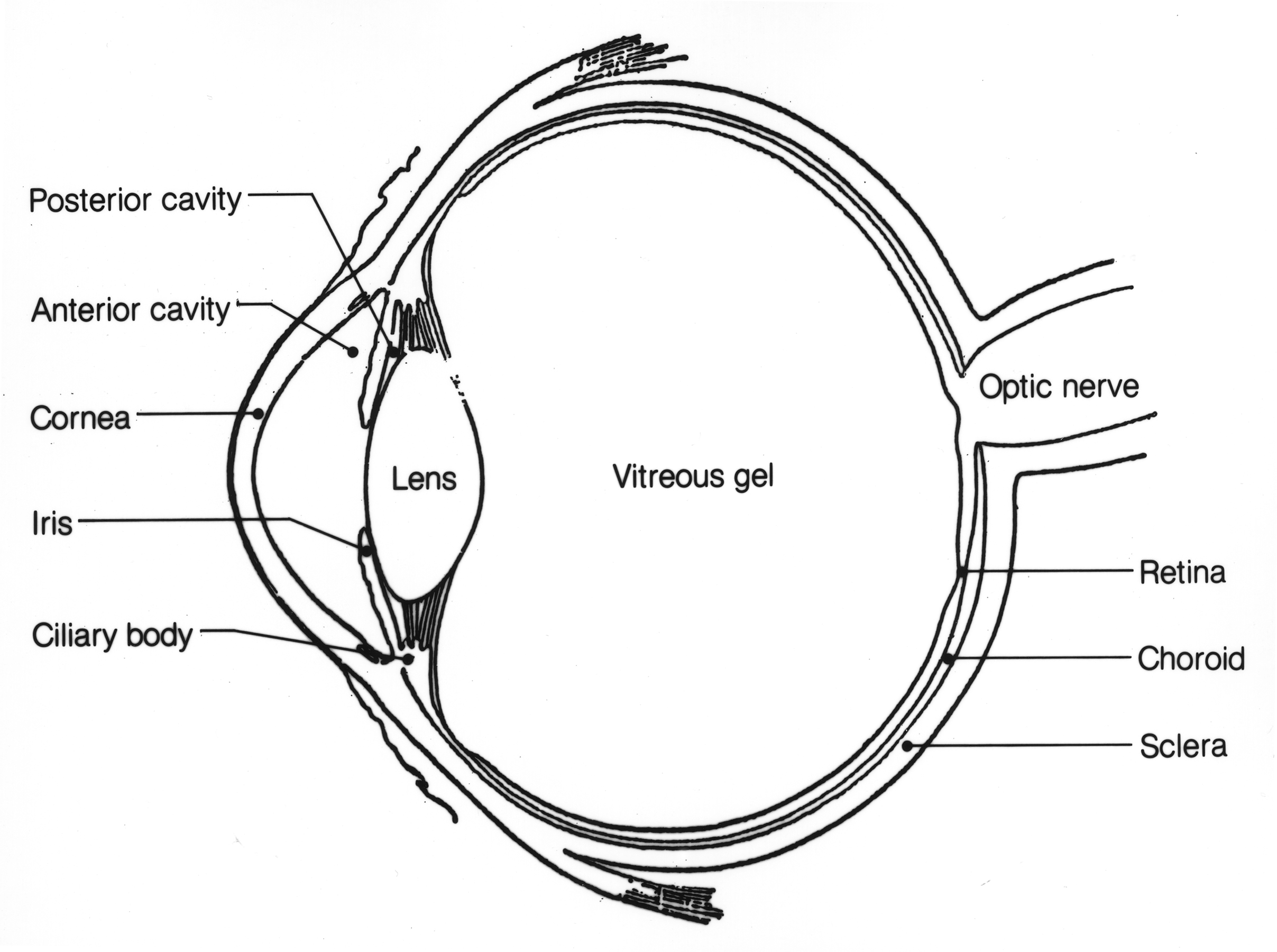
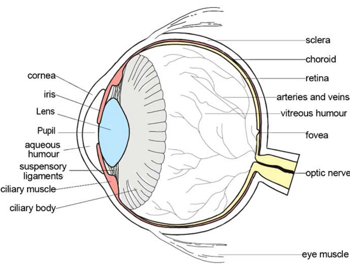
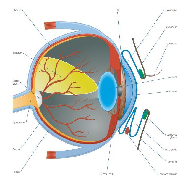
45 diagram of a cow eye
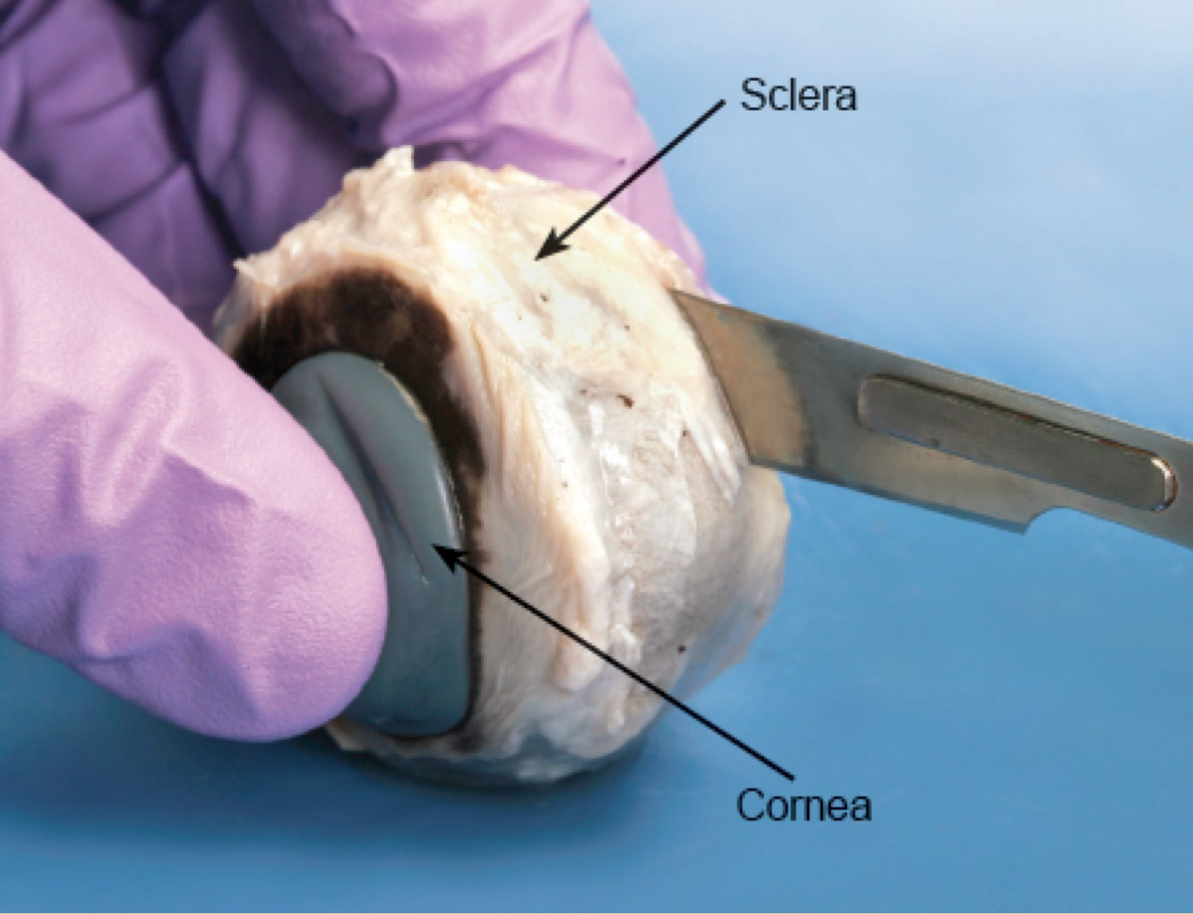
PDF Cow Eye Dissection Guide - cbsd.org DISSECTION OF THE COW EYE Please make sure to wear gloves and safety glasses when you are dissecting, and make sure to clean up thoroughly after the lab. Also, the cow eyes can be rather slippery, so use caution when handling and cutting them. You will need a scalpel and forceps. 1. First, identify the most external structures of the eye. Cow Eye Dissection - The Biology Corner 1. Examine the outside of the eye. You should be able to find the sclera, or the whites of the eye. This tough, outer covering of the eyeball has fat and muscle attached to it 2. Locate the covering over the front of the eye, the cornea. When the cow was alive, the cornea was clear. In your cow's eye, the cornea may be cloudy or blue in color. 2.
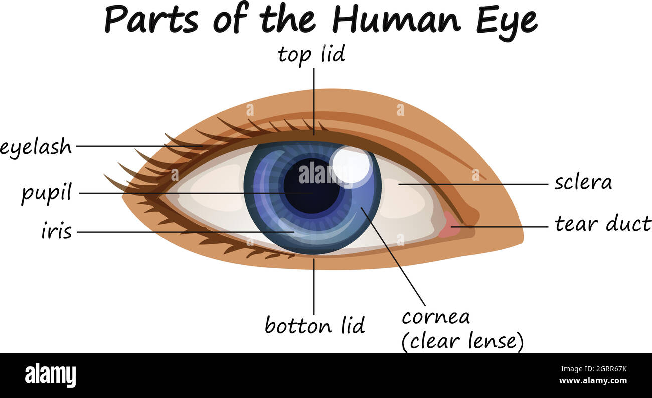
Eye Anatomy: Parts of the Eye and How We See The surface of the eye and the inner surface of the eyelids are covered with a clear membrane called the conjunctiva. The layers of the tear film keep the front of the eye lubricated. Tears lubricate the eye and are made up of three layers. These three layers together are called the tear film. The mucous layer is made by the conjunctiva.
Diagram of a cow eye
Cow's Eye Dissection - step 1 - Exploratorium The blue is the cornea, which starts out clear but becomes cloudy after death. WATCH VIDEO. eye diagram · home · next step. © Exploratorium | The museum ... COW EYE DIAGRAM Quiz - purposegames.com COW EYE DIAGRAM by Robert Stauber8907 3 plays 10 questions ~30 sec English More 0 too few (you: not rated) Tries Unlimited [?] Last Played February 2, 2023 - 05:13 PM There is a printable worksheet available for download here so you can take the quiz with pen and paper. Remaining 0 Correct 0 Wrong 0 Press play! 0% 08:00.0 Highscores Cow Eye Dissection & Parts of the Eye Diagram | Quizlet Transmits visual information from the retina to the brain. tapetum. Give animals night vision; a reflective layer in the eyes of many animals causing them to shine in the dark. cornea. Clear, outer layer of the front of the eye. sclera. White, outermost layer of the eye. Helps maintain shape and gives attachment to muscles.
Diagram of a cow eye. diagram of a cow eye cow eye diagram anatomy labeled exploratorium dissection eyes human edu learning functions parts cows chart studio science section school kids. A guide to beef cuts with steak and roast names : article. Goat anatomy: parts of a goat in english with pictures • 7esl. Cat veins vein diagram jugular internal subscapular thoracic cavity anterior ... Cow's Eye Dissection - Eye diagram - Exploratorium Learn how to dissect a cow's eye in your classroom. This resource includes: a step-by-step, hints and tips, a cow eye primer, and a glossary of terms. Cow's Eye Dissection - Eye diagram The Exploratorium is more than a museum. Explore our online resourcesfor learning at home. © Exploratorium| The museum of science, art and human perception Bull and the cow - Illustrated atlas : normal anatomy | vet ... - IMAIOS Bovine anatomy - Illustrated atlas. This module of vet-Anatomy provides the basics on the anatomy of the bull for students of veterinary medicine. This veterinary anatomical atlas includes 27 scientific illustrations with a selection of labelled structures to understand and discover animal anatomy (skeleton, bones, muscles, joints and viscera). Cow Eye Dissection | Carolina.com Identify the sclera, or tough outer covering of the eyeball. Cow Eye Internal Anatomy Hold the eye between your thumb and forefinger, as shown below. Using scissors or a scalpel, carefully cut the eye in half, separating the front and back of the eye. Examine the inside front portion of the eye. Remove the gelatinous vitreous humor and hard lens.
Cow's Eye Dissection - Eye diagram - Exploratorium A muscle that controls how much light enters the eye. It is suspended between the cornea and the lens. A cow's iris is brown. Human irises come in many colors, ... Equine vision - Wikipedia The eyelids are made up of three layers of tissue: a thin layer of skin, which is covered in hair, a layer of muscles which allow the lid to open and close, and the palpebral conjunctiva, which lies against the eyeball. The opening between the two lids forms the palpebral tissue. The upper eyelid is larger and can move more than the lower lid. Anatomy- Cow Eyeball Diagram | Quizlet clear fluid that helps the cornea keep round shape. Iris. muscle controlling how much light enters the eye. Lens. flexible structure that makes image on retina. Vitreous Humor. thick, clear jelly helps give eyeball shape. Sclera. thick, tough outer covering. Eye Anatomy - 3D model by MotionCow [5dac474] - Sketchfab Vertices: 51.7k. More model information. Realistic cross-section of the Eye. This 3D model can be licensed from MotionCow by Educators, 3D Artists and App Developers. Published 6 years ago. Science & technology 3D Models. eye. cross. anatomy.
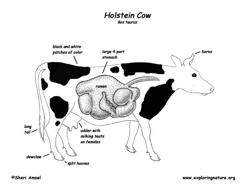
Labeled Cow Eye Diagram - Rock Wiring Cow anatomy diagram showing. Use the point of a scissors or a scalpel to make an incision through the layers of the eye capsule (similar to figure 1); There are three layers from the exterior: Cow Eye Dissection & Labeling Cow eye dissection optic disc pupil cornea sclera nerve iris retina tapetum powerpoint edu ppt presentation. What are the Differences Between a Cow Eye & Human Eye? The radius of the average cow eyeball is a little over 1/2 inch (15mm) with a diameter of 1.2 inches (30mm). The human eyeball size varies, but on average its diameter is about 1 inch (24mm). A cow eyeball and a human eyeball are similar in overall appearance but the parts of the cow eyeball are generally larger than in a human eyeball. Anatomy Eye Structure and Function in Cats - Merck Veterinary Manual Eye, cat. The bony cavity or socket that contains the eyeball is called the orbit. The orbit is a structure that is formed by several bones. The orbit also contains muscles, nerves, blood vessels, and the structures that produce and drain tears. The white of the eye is called the sclera. This is the relatively tough outer layer of the eye. Cow Anatomy - Diagrams Of Cows & Calves - Animal Corner Cow Anatomy Below is a diagram of the Anatomy of a Cow As you can see, there are many parts to a cow. Cows vary in all different colors, some are brown, tanned, white, black, brown-white patched or black-white patched. In a female cow, milk is produced in the udders and extracted from the teats.
Parts and Functions - Dissecting A COW EYE The four muscles that moves the cow's eye in four directions. Cornea A covering over the iris that helps to protect the eye by making light bend to project the image. Iris and Pupil The iris is a muscle that controls how much light goes into the eye and suspended between the cornea and lens. As we have seen in the dissection a cow's iris is brown.
Cow Eye Anatomy Flashcards | Quizlet Cornea Clear protective covering over the front of eye that bends light entering eye Pupil The hole where light passes into the lens Iris The colored part of the eye that regulates the size of the pupil to control light entering the eye Vitreous Humor A clear liquid inside eyeball that gives the eye its round shape Lens
Cow Anatomy - External Body Parts and Internal Organs with Labeled Diagram Other organs systems of cow - you will also learn the anatomical facts of organs from other systems of a cow (showing in diagrams). External body parts of a cow. Okay, you should become familiar with the different external body parts of cow anatomy. This is very simple but important to know the terminologies of cow body parts.
COW'S EYE dissection - Exploratorium cuts the cow’s eye. Whenever you handle raw meat (whether it’s a cow’s eye or a steak), you wash your hands thoroughly afterward to wash away any bacteria you picked up from the meat. If you have cuts on your hand, we also recommend you wear gloves so that no bacteria from the cow’s eye infects your cut. Dissecting a Cow’s Eye
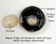
Cow Eye Dissection & Anatomy Project | HST Learning Center Cow Eye Dissection: Internal Anatomy 1. Place the cow’s eye on a dissecting tray. The eye most likely has a thick covering of fat and muscle tissue. Carefully cut away the fat and the muscle. As you get closer to the actual eyeball, you may notice muscles that are attached directly to the sclera and along the optic nerve.
Cow's Eye Dissection | Exploratorium Learn how to dissect a cow's eye in your classroom. This resource includes: a step-by-step, hints and tips, a cow eye primer, and a glossary of terms. Cow's Eye Dissection | Exploratorium The Exploratorium is more than a museum. Explore our online resourcesfor learning at home. Use Policy | Credits ©
COW EYE DIAGRAM — Printable Worksheet COW EYE DIAGRAM — Printable Worksheet Download and print this quiz as a worksheet. You can move the markers directly in the worksheet. This is a printable worksheet made from a PurposeGames Quiz. To play the game online, visit COW EYE DIAGRAM Download Printable Worksheet Please note!
Cow Eye Dissection Teaching Resources | Teachers Pay Teachers by. CrazyScienceLady. 49. $5.00. PDF. Cow Eye Dissection: Directions and questions for dissecting a cow eye. Also includes a diagram of the eye, fun facts, an activity on how to find your blind spot and all answer keys. Appropriate for grades 4-7, I run this lab for each of the 4th grade classes at my school.
Cow's Eye Dissection - How does your eye work? You can do that here. Eye Links · eye diagram · home. © Exploratorium | The museum of science, art ...
Live Science: Cow Eye Dissection - YouTube Watch a Science Museum of Virginia educator conduct our popular cow eye dissection demo.Thanks for watching! Be sure to subscribe to our channel ....
Eye Diagram Basics: Reading, Analyzing and Applying Eye diagrams usually include voltage and time samples of the data acquired at some sample rate below the data rate. In Figure 1 , the bit sequences 011, 001, 100, and 110 are superimposed over one another to obtain the final eye diagram. Figure 1 These diagrams illustrate how an eye diagram is formed. A perfect eye diagram contains an immense ...
Cow Eye Dissection Guide - Google Slides Cow Eye Click Iris: Structure of the eye which controls the size of the opening into the eye which is called the pupil. The pupil gets larger when the radial muscles of the iris contract in...
Cow Eye Dissection Worksheet - BIOLOGY JUNCTION 1. Tell three observations you made when you examined the surface of the eye: 2. Identify the following structures: 3. Name the three layers you sliced through when you cut across the top of the eye: 4. Match the following parts of the eye to their function: (ciliary body, sclera, iris, retina, lens, & tapetum lucidum)
rib eye cut diagram Chart cuts beef meat prime anatomy rib diagram eye ... beef cow roast chuck steak where cuts mignon filet come arm eye pot cooking skirt ribeye mock parts use steaks. The Carnivorous Food Breeze: Smoked Beef Short-Ribs. foodbreeze.blogspot.com. ribs beef short where diagram location cuts cow come rib cut pork prime steak eye country bbq smoked breeze carnivorous. Playing With Fire And Smoke: Guide ...
Beef Cuts Chart and Diagram, with Photos, Names, Recipes, and More Beef Cuts Chart and Diagram, with Photos, Names, Recipes, and More Swap the oven for indirect heat in your grill or smoker to recreate this almost festive flavored recipe: Star anise and orange glazed beef blade roast. Recreate oven temps with indirect heat on your grill for this one: Garlic and tri-pepper-crusted beef roast with balsamic sauce
Cow Eye Dissection & Parts of the Eye Diagram | Quizlet Transmits visual information from the retina to the brain. tapetum. Give animals night vision; a reflective layer in the eyes of many animals causing them to shine in the dark. cornea. Clear, outer layer of the front of the eye. sclera. White, outermost layer of the eye. Helps maintain shape and gives attachment to muscles.
COW EYE DIAGRAM Quiz - purposegames.com COW EYE DIAGRAM by Robert Stauber8907 3 plays 10 questions ~30 sec English More 0 too few (you: not rated) Tries Unlimited [?] Last Played February 2, 2023 - 05:13 PM There is a printable worksheet available for download here so you can take the quiz with pen and paper. Remaining 0 Correct 0 Wrong 0 Press play! 0% 08:00.0 Highscores
Cow's Eye Dissection - step 1 - Exploratorium The blue is the cornea, which starts out clear but becomes cloudy after death. WATCH VIDEO. eye diagram · home · next step. © Exploratorium | The museum ...















Komentar
Posting Komentar