38 sheep brain diagram labeled
Stimulus: Definition, Types, and Examples - Research Tweet 27/08/2021 · • An observable change in the environment is referred to as a stimulus. • Receptors: Environmental inputs are converted into electrical nerve impulses by the receptor. • Neurons: Neurons are the carriers of nerve signals to the central nervous system. • Effectors: As a result of the stimuli, effectors create a reaction. Muscles and glands are examples. Sheep Brain Anatomy and Function Flashcards - Cram.com An extremely efficient memory center in the brain. Responsible for spatial memory and spatial navigation as well as some types of non-spatial memory such as contextual memory and episodic memory. Damage to this structure produces both anterograde and retrograde amnesia. - Vertical cut through pineal gland laterla to pineal gland.
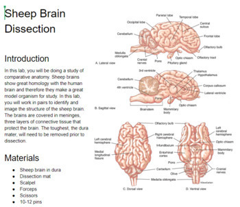
PDF DISSECTION OF THE SHEEP'S BRAIN - Hanover College DISSECTION OF THE SHEEP'S BRAIN Introduction The purpose of the sheep brain dissection is to familiarize you with the three-dimensional structure of the brain and teach you one of the great methods of studying the brain: looking at its structure. One of the great truths of studying biology is the saying that "anatomy precedes physiology".

Sheep brain diagram labeled
Recombinant DNA Technology (With Diagram) - Biology … Biotechnologists have successfully produced transgenic pigs, sheep, rats and cattle. (3) Production of Hormones: By the advent of techniques of rec DNA technology, bacterial cells like E.coli are utilized for the production of different fine chemicals like insulin, somatostatin, somatotropin and p-endorphin. GRADE: 5 SUBJECT: NATURAL SCIENCES AND TECHNOLOGY TERM … B. Sheep C. Lion D. Giraffe. (1) (2) Question 2 Match the definitions in column A with the word in column B. Write the letter from column B as your answer in the middle column. 2. COLUMN A Answer COLUMN B 2.1 Begins to sprout or grow into a seed A. Habitat 2.2 The place where two or more bones meet B. Germination PDF Lab: Sheep Brain Dissection - Mrs. Moretz's Science Site to anatomy studies. See for yourself what the . cerebrum, cerebellum, spinal cord, gray matter, white matter, and other parts of the brain look like! Observation: External Anatomy . 1. You'll need a . preserved sheep brain. for the dissection. Set the brain down so the flatter side, with the white . spinal cord. at one end, rests on the ...
Sheep brain diagram labeled. labeled brain | Brain anatomy, Anatomy and physiology ... - Pinterest Sheep Brain Images taken from the dissection of the sheep's brain: cerebrum, cerebellum, corpus callosum, lobes, sulci, gyrus, fornix, pituitary. H Hillary Hunt 317 followers More information labeled brain Find this Pin and more on NeuroBiology by Hillary Hunt. Brain Anatomy Human Anatomy And Physiology Medical Anatomy Medical Student Study Sheep Brain Anatomy Quiz - ProProfs Quiz 1. What is the outer covering of the Sheep brain? A. Arachnoid Mater B. Pia Mater C. Medulla Oblongata D. Dura Mater 2. Which amongst the following is the largest structure in dorsal view? A. Pons B. Medulla Oblongata C. Cerebral cortex D. Hippocampus 3. What are the large folds that surround the cerebrum? A. Gyri B. Sulci C. Fissures D. Colliculus Sheep Brain - San Diego Mesa College Sheep Brain. Click on a photo for a larger view of the model. Click on Label for the labeled model. Back to Dissected Specimen Page. Superior View: Corpora Quadrigemina: Label: Label: Sagittal Section -lateral ventricle: Sagittal Section - septum pellucidum: Label: Label: Inferior View: Coronal Section: Clitoris - Wikipedia The clitoris (/ ˈ k l ɪ t ər ɪ s / or / k l ɪ ˈ t ɔːr ɪ s / ()) is a female sex organ present in mammals, ostriches and a limited number of other animals.In humans, the visible portion – the glans – is at the front junction of the labia minora (inner lips), above the opening of the urethra.Unlike the penis, the male homologue (equivalent) to the clitoris, it usually does not ...
Sheep brain labeling Quiz - PurposeGames.com This is an online quiz called Sheep brain labeling Your Skills & Rank Total Points 0 Get started! Today's Rank -- 0 Today 's Points One of us! Game Points 25 You need to get 100% to score the 25 points available Add to Playlist Add to tournament Give a nod to the game author Games by same creator Brachial Plexus 8p 10p Image Quiz 17p Sheep Brain - Dorsal View - University of Minnesota Dissected Sheep Brain — Dorsal View Dorsal view of sheep brain with the cerebellum and caudal cerebrum removed. The rostral colliculus(large arrow label) and the caudal colliculus(small arrow label) together form the tectumof the midbrain. Sheep Brain Label - The Biology Corner Students can practice naming the parts of the brain, then check their answers with the provided key. ... Label theBrain of the Sheep. Publisher: Biologycorner.com; follow on Google+ This work is licensed under a Creative Commons Attribution-NonCommercial 3.0 Unported License. Brain Label Answer Key. Image adapted from a photograph of the sheep ... Cow Anatomy – External Body Parts and Internal Organs with Labeled Diagram 28/07/2021 · Why are the sheep brain and cow eye used to study anatomy? Why are cow hearts used to study human anatomy? Conclusion. This is the basic information on cow anatomy for any veterinary student or farm owner. But, you might learn more about different systems of cow anatomy with a labeled diagram. , you may visit the general anatomy section of ...
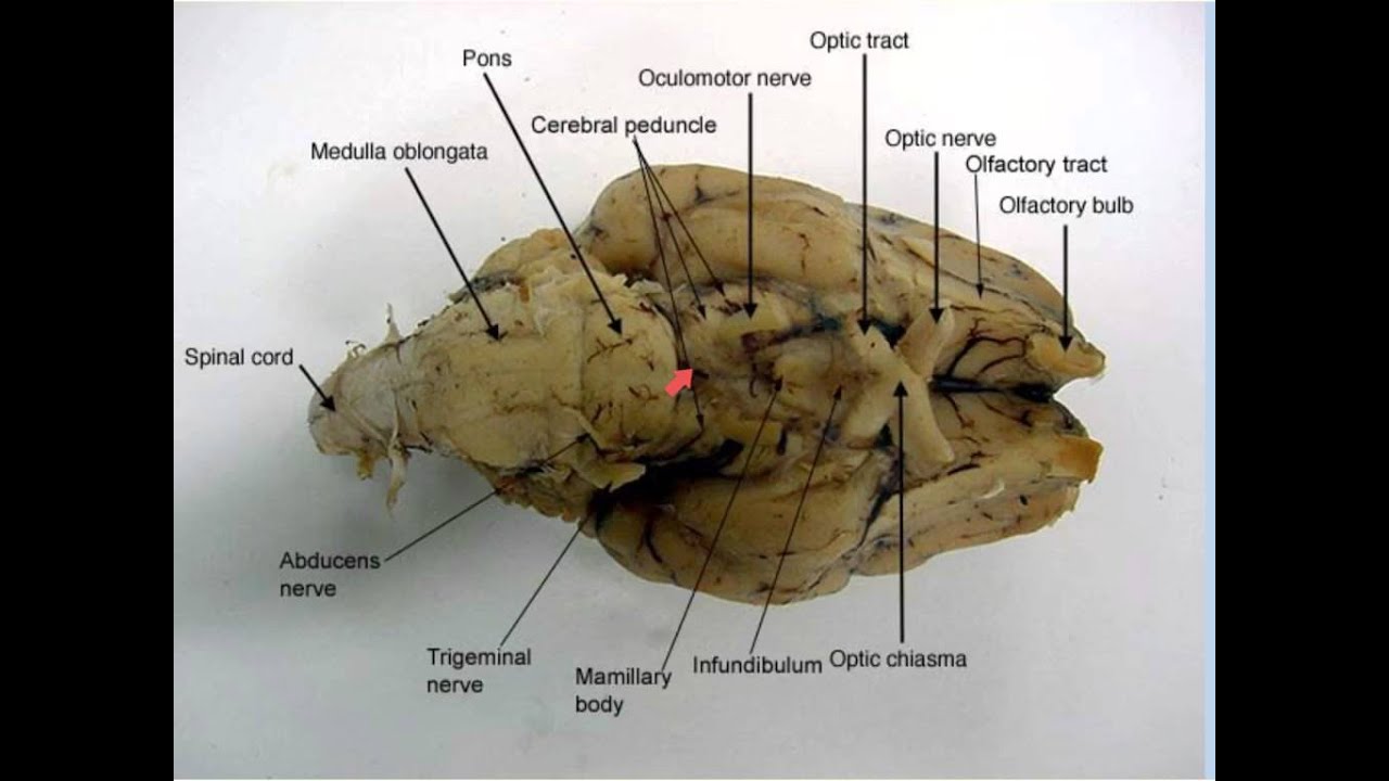
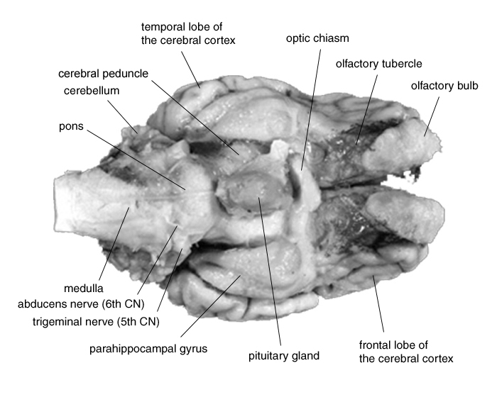
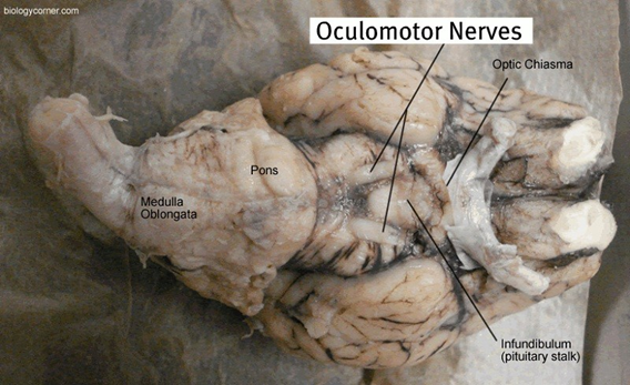
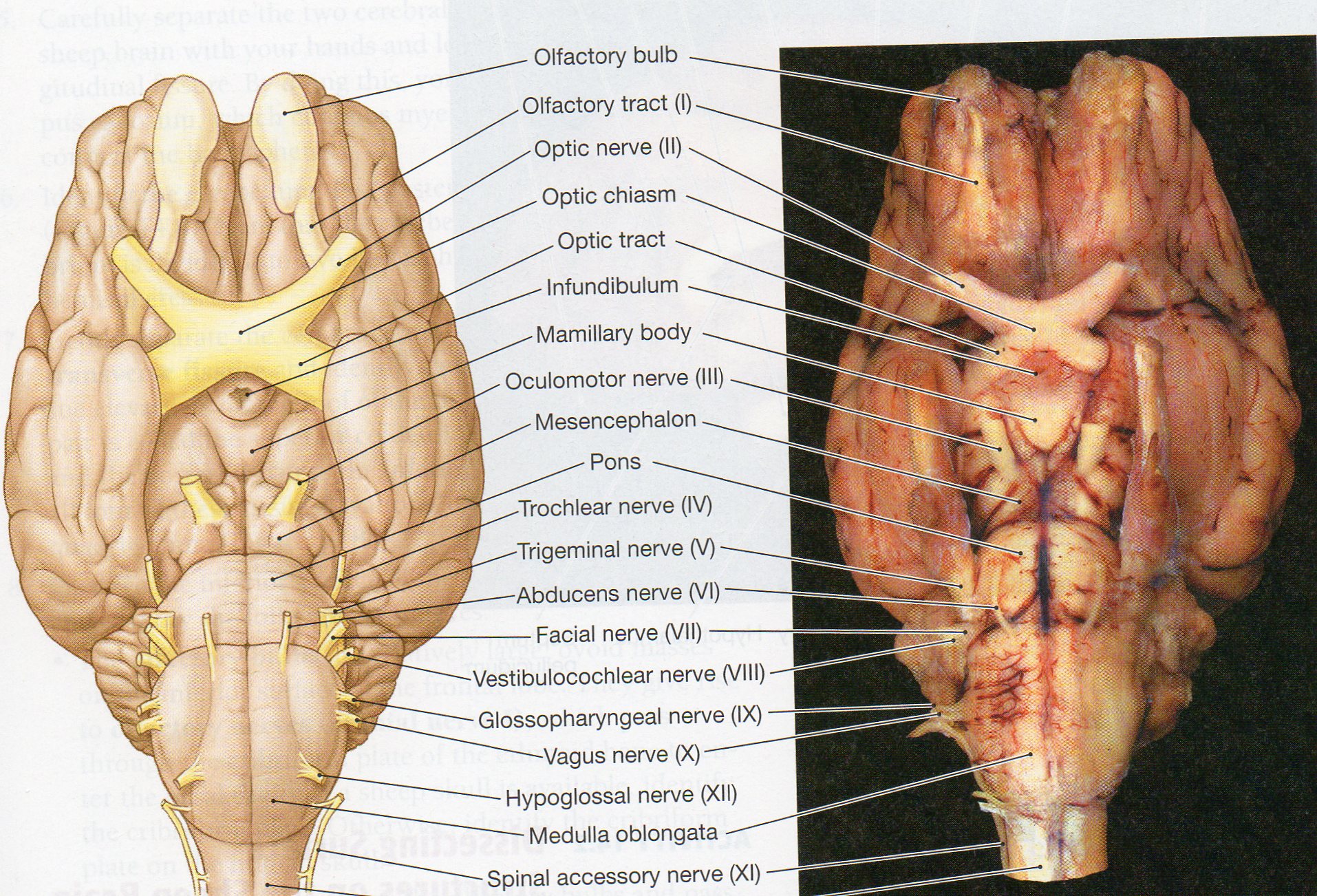
Sheep Brain external view labeled | Brain anatomy, Feline anatomy, Brain A & P. Nerve Anatomy. Anatomy And Physiology. Neuron Diagram. Brain Science. Science Biology. Pig Brain. 1.1 - Identify the role of receptors in detecting stimuli. Stimuli are environmental factors that can be either internal or external, these factors trigger a response and the organism reacts... learning-center.homesciencetools.com › articleSheep Heart Dissection Lab for High School Science | HST By studying the sheep’s anatomy, you can learn how your own heart pumps blood through your body, thereby keeping you alive! Use this sheep heart dissection guide in a lab for high school students. You can also look at the labeled pictures to get an idea of what the heart looks like (that’s especially helpful for younger students). Sheep brain dissection | Human Anatomy and Physiology Lab (BSB 141 ... The most prominent structure visible on the ventral side of the sheep brain is half of the optic chiasma, which is where the two optic nerves cross over each other and form an "X" shape. You will only see half the structure. Find the optic chiasma half on your brain. You may have removed the optic removed the chiasma with the dura mater. Hypothalamus - Wikipedia The hypothalamus (from Ancient Greek ὑπό (hupó) 'under', and θάλαμος (thálamos) 'chamber') is a part of the brain that contains a number of small nuclei with a variety of functions. One of the most important functions is to link the nervous system to the endocrine system via the pituitary gland.The hypothalamus is located below the thalamus and is part of the limbic system.
File:Sheep Brain Dissection 2 - black background.png - Wikipedia
PDF Neuroanatomy: Dissection of the Sheep Brain - Napa Valley College Examine the sheep brain with the membranes intact. You should be able to identify and use the following directional terms: Anterior / Posteriorfront / back Rostral / Caudal towards the beak / towards the tail Medial / Lateral towards the middle / towards the side Dorsal / Ventral top / bottom (on the CNS of a quadruped)
sheep brain anatomy
Sheep Brain Dissection Project Guide | HST Learning Center Use the labeled picture to identify the corpus callosum, medulla, pons, midbrain, and the place where the pituitary gland attaches to the brain. (In many preserved specimens the pituitary gland is no longer present. It is not pictured.) Use your fingers or a teasing needle to gently probe the parts and see how they are connected to each other.
Final Brain Basics-1
Spongiform Encephalopathy in Transgenic Mice Expressing a Point ... 09/09/2011 · A, Soluble (S) and insoluble (P) PrP from brain homogenate were separated by ultracentrifugation and immunoblotting. Percentage of PrP in the pellet fractions was quantified and graphed, with each point representing an individual mouse. Samples labeled L81 yo were from 40-d-old mice, where as all other uninoculated mice were older than 300 d.

Sheep Brain external view labeled | Brain anatomy, Feline ...
› dna › recombinant-dnaRecombinant DNA Technology (With Diagram) - Biology Discussion Biotechnologists have successfully produced transgenic pigs, sheep, rats and cattle. (3) Production of Hormones: By the advent of techniques of rec DNA technology, bacterial cells like E.coli are utilized for the production of different fine chemicals like insulin, somatostatin, somatotropin and p-endorphin.
Sheep Brain Dissection Guide
Segmentation: U-Net, Mask R-CNN, and Medical Applications 21/01/2020 · (d) instance segmentation, in which the model assigns an “individual object” label to each pixel in the image. In this example, the pixels for each individual sheep are labeled separately. Instead of having a generic “sheep” pixel class, we now have five classes for the five sheep shown: sheep1, sheep2, sheep3, sheep4, and sheep5.
Sheep Brain Dissection
11.7: Sheep Brain Dissection - Biology LibreTexts Background Information: The sheep brain is remarkably similar to the human brain. One major difference, however, is in proportion. For example, the sheep brain has a proportionately smaller cerebrum. Another difference is in orientation of the spinal cord. The sheep spinal cord is orientated anterior to posterior, as in any four-legged animal.
Sheep Brain Explora on Guide
Sheep Brain Dissection Lab Use the picture below as a way to see how your sheep brain should look after you cut it in half. 10. In the image below, a probe indicates the location of the lateral ventricle. 11. Once the brain is cut this way, the colliculi can also be seen from the inside and the pineal gland is revealed only if you made a very careful incision.
Lab - Sheep Brain: MAH-Summer 2019-Anatomy and Physiology I
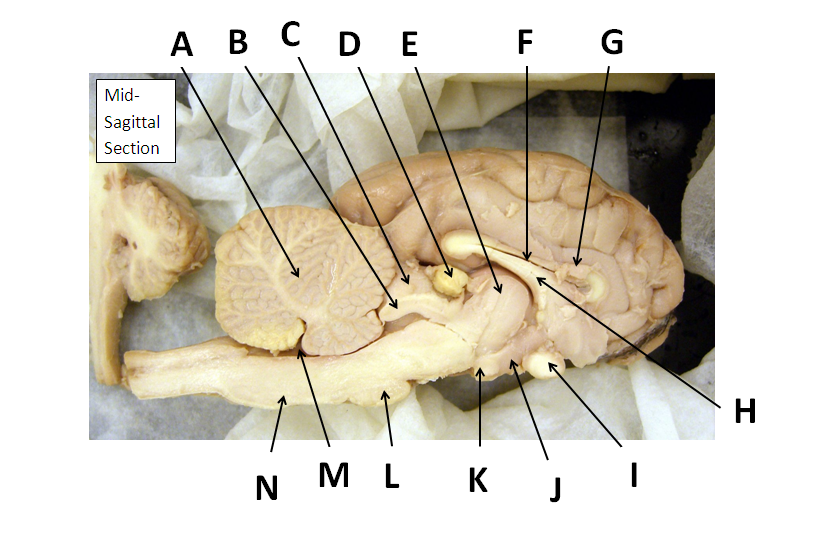
Solved Art-labeling Activity: Midsagittal Section of the - Chegg Anatomy and Physiology questions and answers. Art-labeling Activity: Midsagittal Section of the Sheep Brain (Diagram, 2 of 2) Reset Help Fomix Infundibulum Olfactory bulb Optic chiasm Mosencephalon Pituitary gland Marillary body Medulla oblongata Pons Spinal cord Corpus callosum Art-labeling Activity: Midsagittal Section of the Sheep Brain ...
The Brain Biodiversity Bank at Michigan State University
brain parts labelled diagram - Microsoft brain sheep label anatomy labeled superior classroom labels sdmesa physiology nervous inferior edu lateral sup ventricle section sagittal creative coronal Brain anatomy function lobe sagittal lateral frontal occipital normal medicalartworks temporal parietal anatomical psychology neuroscience. Dog eye anatomy. Brain stem and adjacent structures
Sheep Brain
PDF Sheep Brain Dissection Lab - Home Science Tools Use the labeled picture to identify the corpus callosum, medulla, pons, midbrain, and the place where pituitary gland attaches to the brain. (In many preserved specimens the pituitary gland is no longer present. It is not pictured.) Use your fingers or a teasing needle to gently probe the parts and see how they are connected to each other.

Sheep brain | Atlas of Comparative Vertebrate Anatomy
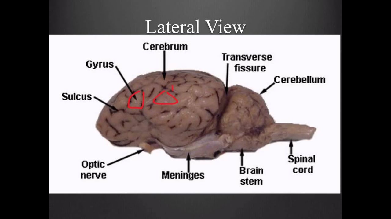
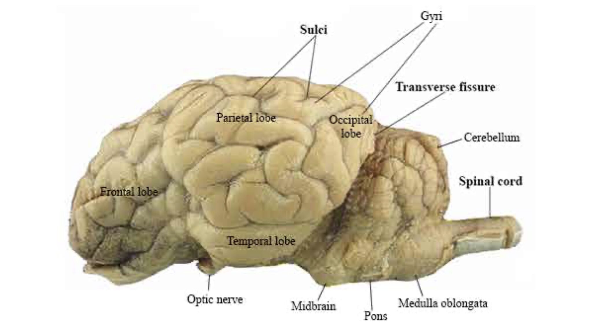
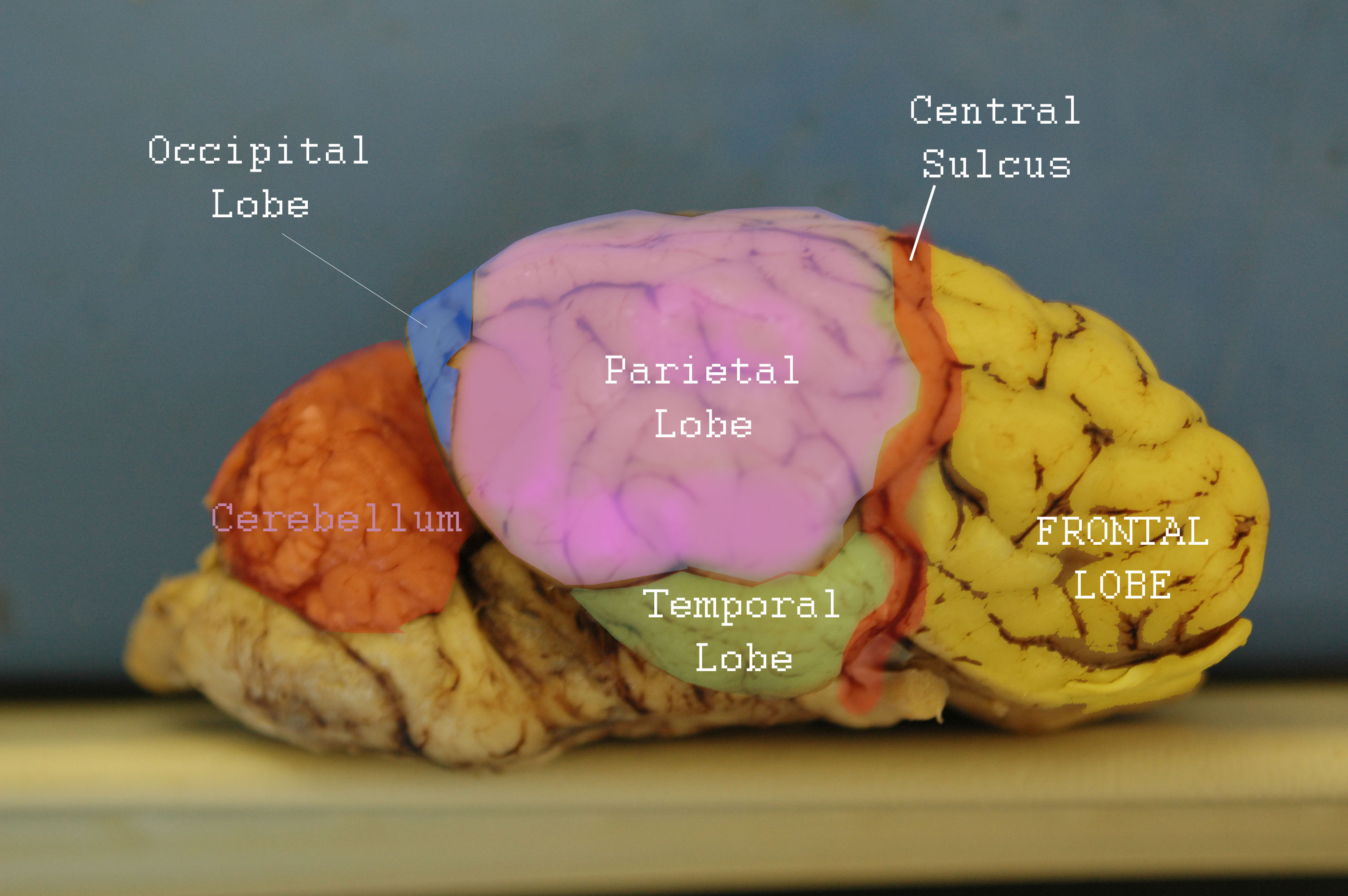
DOC Sheep Brain Anatomy Lab Manual - amherst.edu The cruciate fissure (labeled ansate sulcus in your photo atlas) is known in the human brain as the fissure of Rolando or central sulcus, and intersects the medial longitudinal fissure to mark off the anterior third of the cortex.
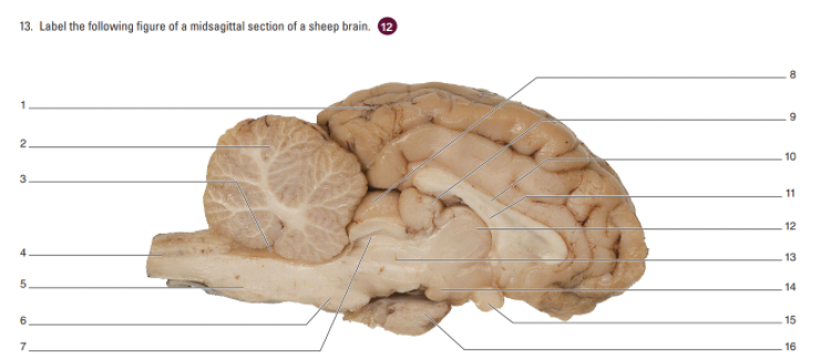
Solved Label the following figure of a midsagittal section ...
en.wikipedia.org › wiki › HypothalamusHypothalamus - Wikipedia Fos-labeled cell analysis showed that the PMDvl is the most activated structure in the hypothalamus, and inactivation with muscimol prior to exposure to the context abolishes the defensive behavior. Therefore, the hypothalamus, mainly the PMDvl, has an important role in expression of innate and conditioned defensive behaviors to a predator.

Sheep brain quiz Flashcards | Quizlet
Sheep Brain Neuroanatomy Online Self-Test | KPU.ca - Kwantlen ... Use each diagram as a reference, and selected the correct answer for each lettered structure. ... Sheep Brain Neuroanatomy Online Self-Test. Use each diagram as a reference, and selected the correct answer for each lettered structure. You may find it useful to open the diagrams in a separate window to review while answering each question.
11.7: Sheep Brain Dissection - Biology LibreTexts
researchtweet.com › stimulus-definition-types-andStimulus: Definition, Types, and Examples - Research Tweet Aug 27, 2021 · The arteries stretch when blood pressure is too high, and receptors transmit a signal to the brain. The heart rate will be lowered by the brain. The absence of a signal from the receptor indicates that the blood pressure is low. To maintain normal blood pressure, the brain will raise the heart rate.
1 Dissection of the Sheep Brain Applications
Male anatomy drawing labeled 2022. 7. 1. · Anatomical Drawings Of A Fetal Pig blog.valdosta.edu. pig fetal drawing heart anatomy vessels labeled internal diagram blood dissection drawings identify following use male anatomical important paintingvalley. Duke Anatomy - Lab 2 Optional Exercise web.duke.edu. anatomy surface duke optional exercise lab atlas. Dissection Of The Fetal Pig. Line Drawing Of …

Cerebrum Sheep Dissection - Human Anatomy - GUWS Medical
wvrxmq.diekosmonauten.de › male-anatomy-drawingMale anatomy drawing labeled DON’T draw every. brain diagram unlabeled human half anatomy clipart blank left cliparts clip labeled psych ap results etc clipartmag library usf edu. Human Brain And Nervous System - Anatomy 3D Model From CreativeCrash . brain 3d human nervous system organs body anatomy internal skeleton male .
Sheep+brain+dissection
Label Sheep Brain Diagram | Quizlet Label Sheep Brain. STUDY. Learn. Flashcards. Write. Spell. Test. PLAY. Match. Gravity. Created by. telaneyn. Terms in this set (11) Cerebellum. the "little brain" at the rear of the brainstem; functions include processing sensory input and coordinating movement output and balance. Occipital lobe. A region of the cerebral cortex that processes ...
SCB209 - Lab2 - Natural Sciences Open Educational Resources
Sheep Brain Dissection Guide - YouTube About Press Copyright Contact us Creators Advertise Developers Terms Privacy Policy & Safety How YouTube works Test new features Press Copyright Contact us Creators ...
Physiological Psychology
Sheep Brain Label | Dissection, Human brain diagram, Brain anatomy Sheep Brain Label | Dissection, Human brain diagram, Brain anatomy From biologycorner.com Sheep Brain Label A drawing of the brain with the parts unlabeled. Students can practice naming the parts of the brain, then check their answers with the provided key. Biologycorner 17k followers More information unlabeled brain
Sheep Brain Neuroanatomy Online Self-Test | KPU.ca - Kwantlen ...
sheep brain labeled Diagram | Quizlet sheep brain labeled STUDY Learn Write Test PLAY Match + − Created by ARCELIAWALKER PLUS Terms in this set (18) superior colliculus ... pineal gland ... fornix ... corpus callosum ... cerebrum ... septum pellucidum ... optic chiasma ... hypotalamus ... thalamus ... cerebral aqueduct ... cerebral penducle ... mamillary body ... ponds ...
Sheep Brain Neuroanatomy Online Self-Test | KPU.ca - Kwantlen ...
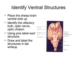
Sheep Brain Dissection with Labeled Images - The Biology Corner 1. The sheep brain is enclosed in a tough outer covering called the dura mater. You can still see some structures on the brain before you remove the dura mater. Take special note of the pituitary gland and the optic chiasma. These two structures will likely be pulled off when you remove the dura mater. Brain with Dura Mater Intact
Index of /files/OCC_VIDEO/upload/Faculty_Resources/acamilo ...
sheep brain worksheet brain anatomy sheep dissection human cerebellum labeled identify parts diagram guide spinal cord physiology cow cerebrum labels corpus callosum nerve. Sheep Brain Dissection Guide With Pictures . dissection labeled homesciencetools. Observation 1 Sketch Of Sheep Brain 3 4 Closely Examine The Cerebrum ...

sheep brain by Melanie Brawley - Issuu
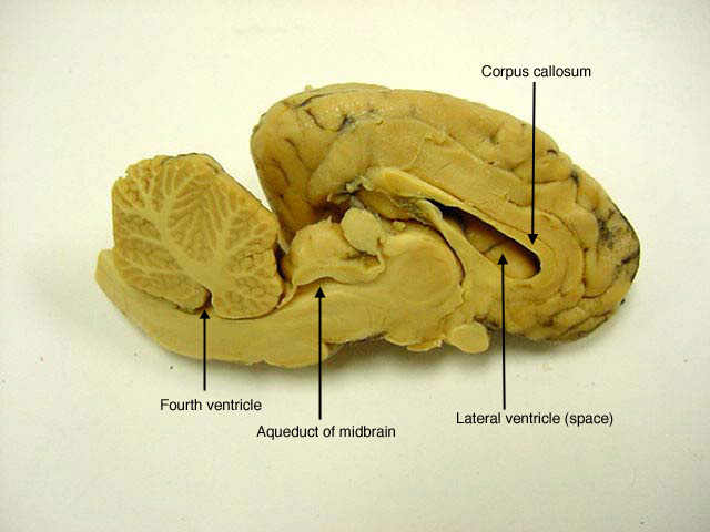
PDF Sheep Brain Midsagittal Section - drcroes.com Sheep Brain -Parasagittal Section 1. Gray Matter 2. White Matter 3. Corpus Callosum 4. Lateral Ventricle 5. Caudate Nucleus 6. Septum Pellucidum 7. Fornix 8. Optic Chiasma 9. Third Ventricle 10. Thalamus (Ovid Nuclear Mass of Thalamus) 11. Corona Radiata 12. Hippocampus 13. Cerebral Aqueduct (of Sylvius) 14. Pituitary Gland (hypophysis) 15.
CENTRAL NERVOUS SYSTEM
Sheep Brain Dissection labeled Diagram | Quizlet Only $2.99/month Sheep Brain Dissection labeled STUDY Learn Write Test PLAY Match Created by AllieKlinger Terms in this set (8) Corpus Collosum Lateral Ventricle Fornix Hypothalamus Cerebral Aqueduct Central Canal Inferior Collicuious Transverse Fissure THIS SET IS OFTEN IN FOLDERS WITH... Sheep Brain Dissection labeled 2 8 terms AllieKlinger
Sheep Brain Dissection Lab
Sheep Heart Dissection Lab for High School Science | HST By studying the sheep’s anatomy, you can learn how your own heart pumps blood through your body, thereby keeping you alive! Use this sheep heart dissection guide in a lab for high school students. You can also look at the labeled pictures to get an idea of what the heart looks like (that’s especially helpful for younger students).
Sheep Brain Dissection Guide
anatomylearner.com › cow-anatomyCow Anatomy - External Body Parts and Internal Organs with ... Jul 28, 2021 · Cow anatomy labeled diagram. Here I would like to summarize the whole anatomical features of a cow (both internal and external) with the labeled diagram. I hope you will enjoy it and learn the anatomical features of the different organs of a cow. If you need more cow-labeled diagrams, you may join with anatomy learners on social media.
Lab: Sheep Brain Dissection
Practice Lab Practical on the Sheep Brain - PGCC Identify the lobe labeled 1. Identify the lobe labeled 2. Identify the lobe labeled 3. Identify the structure labeled 4. Identify the structure lobe labeled 5. ... Identify the shiny membrane visible on the sheep brain surface. In the above picture: Identify the structure labeled 1. Identify the structure labeled 2. ...
Brain Anatomy Labeled Diagram Stock Vector - Illustration of ...
en.wikipedia.org › wiki › ClitorisClitoris - Wikipedia The clitoris (/ ˈ k l ɪ t ər ɪ s / or / k l ɪ ˈ t ɔːr ɪ s / ()) is a female sex organ present in mammals, ostriches and a limited number of other animals.In humans, the visible portion – the glans – is at the front junction of the labia minora (inner lips), above the opening of the urethra.
Lab 12 - The Brain and Cranial Nerves
PDF Lab: Sheep Brain Dissection - Mrs. Moretz's Science Site to anatomy studies. See for yourself what the . cerebrum, cerebellum, spinal cord, gray matter, white matter, and other parts of the brain look like! Observation: External Anatomy . 1. You'll need a . preserved sheep brain. for the dissection. Set the brain down so the flatter side, with the white . spinal cord. at one end, rests on the ...
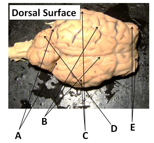
Sheep Neuroanatomy Lab- Labeling Worksheet Figure 1: Dorsal view
GRADE: 5 SUBJECT: NATURAL SCIENCES AND TECHNOLOGY TERM … B. Sheep C. Lion D. Giraffe. (1) (2) Question 2 Match the definitions in column A with the word in column B. Write the letter from column B as your answer in the middle column. 2. COLUMN A Answer COLUMN B 2.1 Begins to sprout or grow into a seed A. Habitat 2.2 The place where two or more bones meet B. Germination

Neuron/Spinal Cord Histology Brain Anatomy Sheep Brain ...
Recombinant DNA Technology (With Diagram) - Biology … Biotechnologists have successfully produced transgenic pigs, sheep, rats and cattle. (3) Production of Hormones: By the advent of techniques of rec DNA technology, bacterial cells like E.coli are utilized for the production of different fine chemicals like insulin, somatostatin, somatotropin and p-endorphin.
A&P 2 Lab page 7

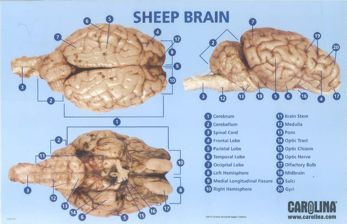
Sheep Brain Dissection | Carolina.com

Sheep Brain
Sheep brain dissection - Bisc 163 - StuDocu
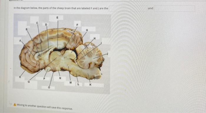
Solved in the diagram below, the parts of the sheep brain ...

Alila Medical Media | Blood supply of the brain, labeled ...

SheepBrain-section-names.jpg? ...


Komentar
Posting Komentar